

WeFixDMD Equipment Store
X-ERA Dental Cone Beam System
Couldn't load pickup availability
For dental professionals that need both 3D volumes AND 2D panoramic radiographs to be high resolution.
Most dental cone beam systems use the same sensor for 2D panoramic radiographs as they use for the 3D scans. This means that one (or both) modality is typically suboptimal.
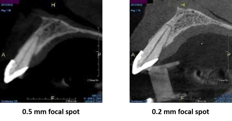
A 0.2 mm focal spot will allow you to spend less time convincing patients of your diagnosis and more time practicing dentistry.
Most cone beam systems either have a 0.5 mm or worse (larger focal spots equal less edge sharpness). The X-era focal spot is one of the smallest in the industry for a dental cone beam system. This not only means you and your staff can see more, but your patients can as well.
Field of View options to go as small as you want, or as big as you need.
Many dental cone beam systems max out at 9 or 10 cm wide. This may lead to:
- Cutting off 3rd molars
- Inability to perform airway analysis
- Inability to capture TMJs
- Little margin for error in patient positioning (i.e. suboptimal positioning may cause desired anatomy to fall outside the scanning volume which may require a retake)
The X-era gives the operator a number of options ranging from endo (4 cm x 6 cm) all way up to 16 cm wide. This offers fantastic flexibility and won’t limit what you can do with the platform.
Low-dose options allow cone beam scans with lower radiation than traditional intraoral exams.
Traditional FMX studies can be 150 microsieverts or more of radiation for the patients. The low-dose modes of the X-era can provide a cone beam scan with less radiation while opening up a world of new information.
A Panoramic X-Ray to be proud of
Most of our dental professionals depend on their cone beam system to produce a high-quality panoramic x-ray in addition to a high-quality 3D volume. Most dental cone beam systems sacrifice the performance of their panoramic modality by attempting to use the 3D sensor to perform 2D panos. The X-era uses a cone beam sensor for cone beam scans, and a panoramic sensor for panoramic scans.
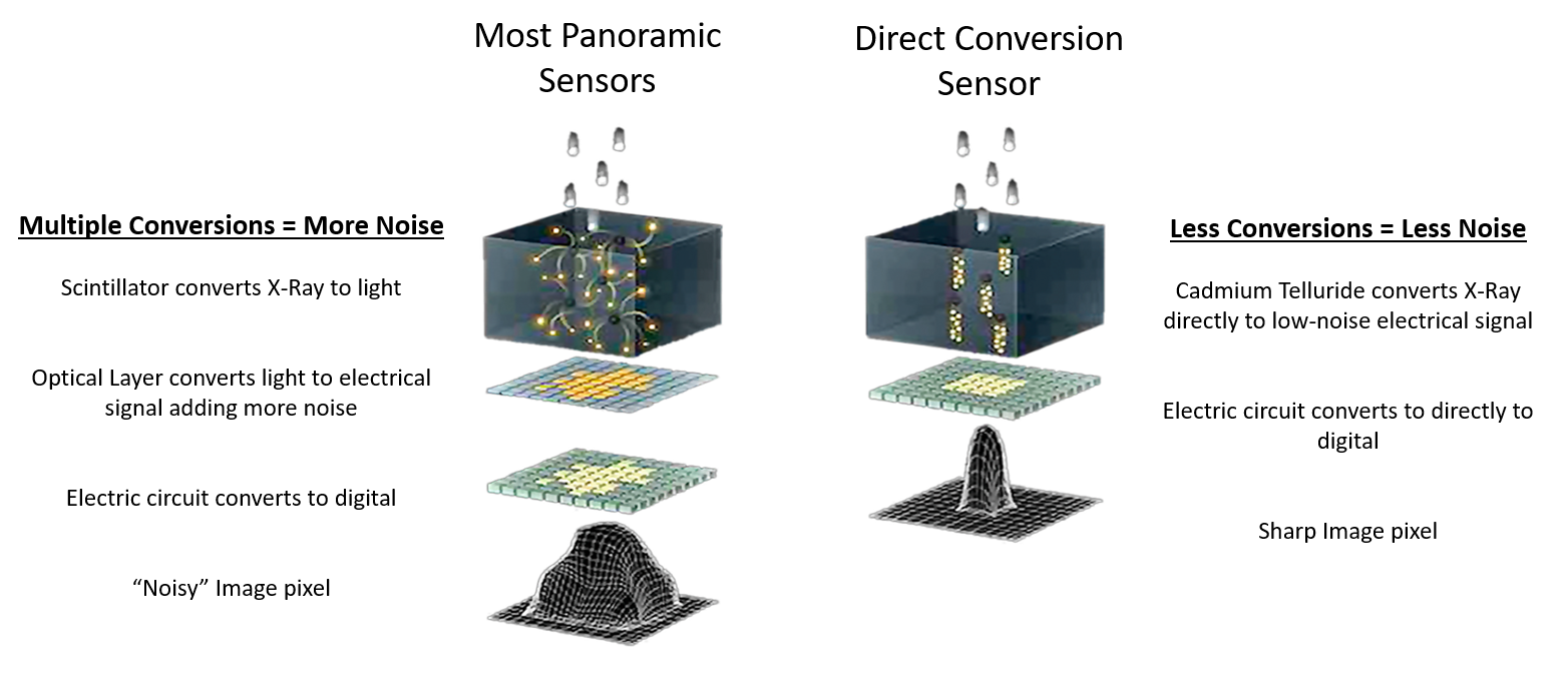
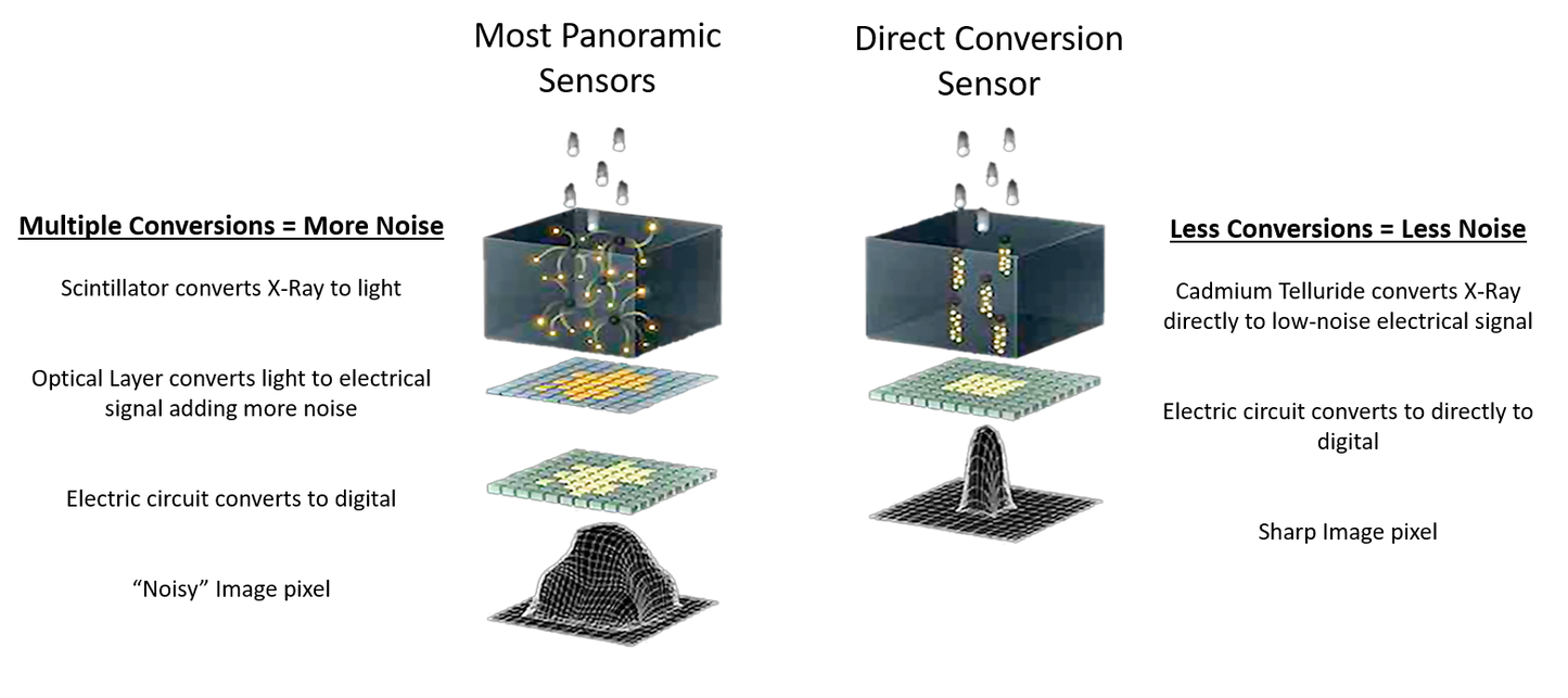
What’s so great about a Direct Conversion Sensor?
Most Panoramic CMOS sensors use lower-cost materials that are less sensitive to x-ray. Therefore, these sensors must convert x-ray to light before converting to a digital signal. This can result in image blurriness because the extra conversion step can cause the radiation to “fan out” and inadvertently trigger surrounding pixels. The X-era utilizes a Direct Conversion (sometimes referred to as “Cadmium Telluride”) sensor that converts x-ray directly to digital, which contributes to a much sharper image.
Every scan captures over 50 different panoramic layers!
With most panoramic x-ray units, the arch of the patient needs to be exactly where the machine wants: along the path where the beam is focused.
- If positioning is not precise, a poor image can result.
- If the patient shifts slightly during the time the operator walks out to initiate the scan, a poor image can result.
- If the patient’s arch doesn’t match the trajectory of the scan, certain anatomy may not be in focus.
- The emergence profile of the anteriors could make it difficult to capture both mandibular and maxillary apices clearly in the panoramic scan.
The X-era also has Multi-Focal Plane capture capability. This means that every scan captures many layers both forward and backward. This creates a wide “envelope” where much more data is captured around where the patient is positioned.
Not only Bitewings, but an entire FMX!
-
Vertical bitewing feature as well as FMX Clipping feature creates 18 intraoral-sized images clipped from the pan with one click.
-
Ideal tool for patients who won’t tolerate traditional PAs
- Instead of taking 18 individual images with an intraoral X-Ray, they can all be generated at once in 14 seconds.
-
Free Shipping
-
Hassle-Free Exchanges